
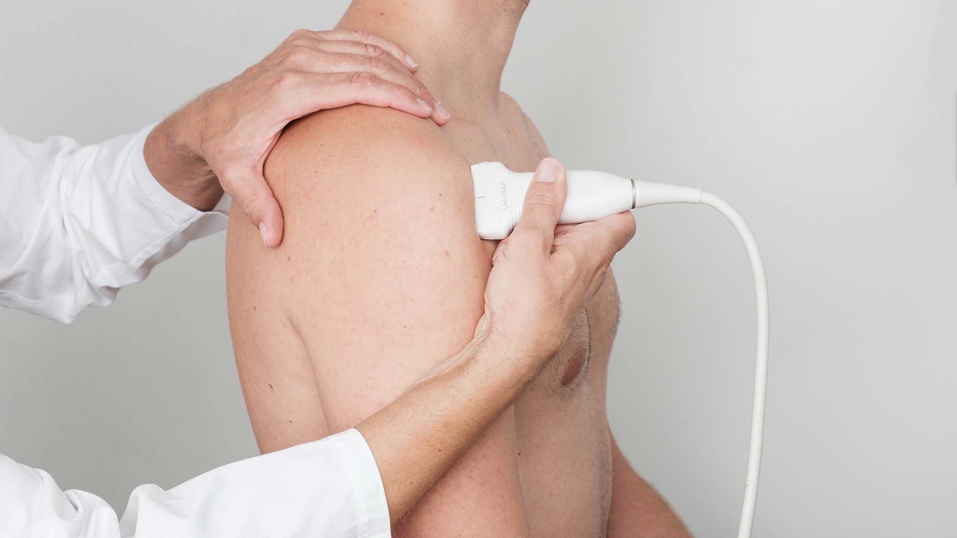
Shoulder ultrasound
Diagnostic shoulder ultrasound is a common diagnostic procedure. The shoulder has plenty of soft tissue near the surface, with bone beneath it, which makes it well suited to ultrasound assessment. The structures of the shoulder interact in a very complex way. The group of small muscles that rotate the shoulder joint is called the rotator cuff; it is made up of the supraspinatus, subscapularis, infraspinatus and teres minor. Other muscles cover them and move the shoulder as well as the forearm (deltoid, biceps, pectoralis). The shoulder has many fluid-filled sacs (bursae), the largest of which is the subacromial-subdeltoid bursa.
Diagnostic ultrasound can detect partial or complete muscle tears, inflammation (tendinitis), degeneration (tendinosis), atrophy, calcifications, impingement syndromes and frozen shoulder (adhesive capsulitis). The bony structures of the shoulder are less accessible, so only surface irregularities can be seen with this type of examination. For everything else, a plain X-ray, computed tomography or magnetic resonance imaging is needed. In inflammatory conditions, laboratory tests are also needed. The shoulder girdle is a complex joint consisting of the glenohumeral, acromioclavicular, sternoclavicular and scapulothoracic joints.
Diagnostic shoulder ultrasound can detect and monitor changes, and on the basis of this work-up various interventional procedures can be carried out, such as injections, pain blocks, instillations, tendon fenestration, dry needling and acupuncture, or various medicines can be given, such as corticosteroids, anaesthetics, hyaluronic acid, platelet-rich plasma, whole blood, stem cells and others. Dynamic evaluation – a dynamic ultrasound examination of the shoulder – makes it possible to see the condition and behaviour of the shoulder structures in real time. Subluxations and dislocations of tendons are therefore sometimes easier to spot. Diagnostic ultrasound is an excellent method for monitoring patients before and after surgery.
The test should always be interpreted in the context of the clinical findings and other diagnostic tests. An examination by a specialist in musculoskeletal disorders, together with an additional diagnostic ultrasound examination, leads to an accurate diagnosis.


