
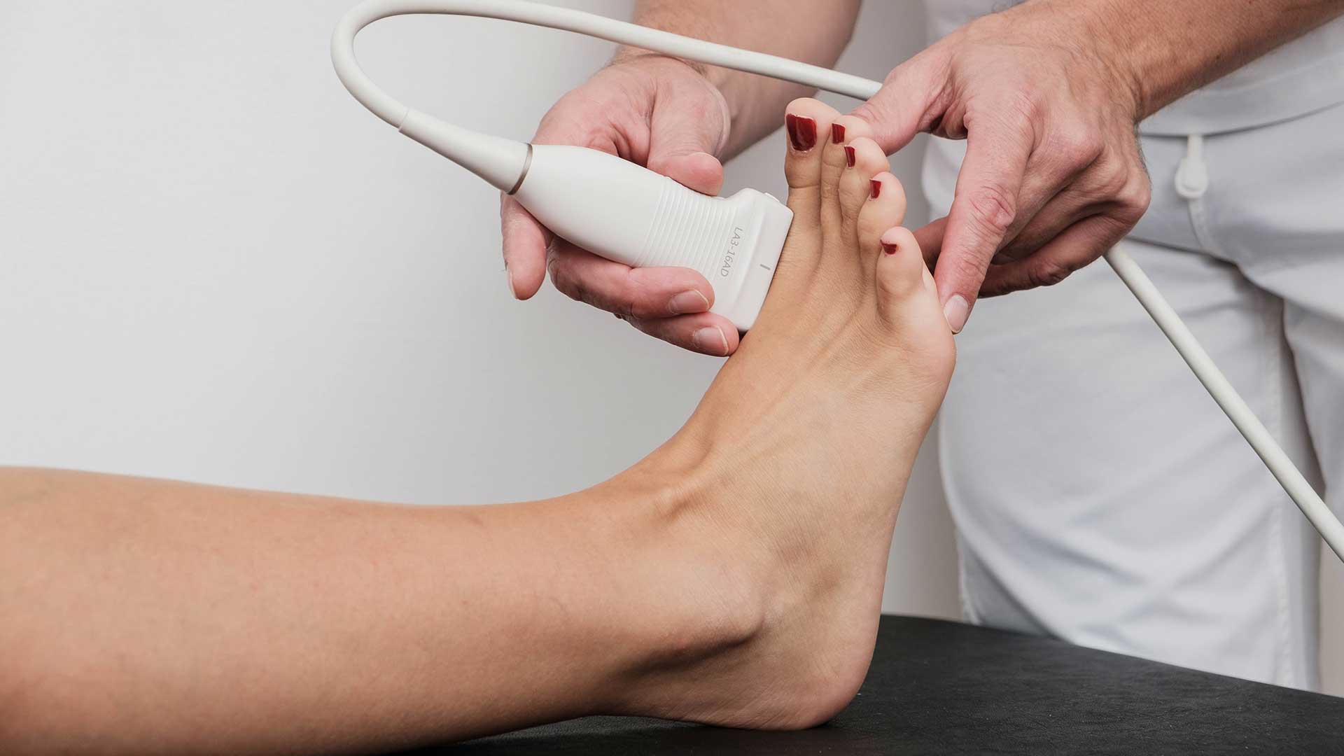
Foot ultrasound
Diagnostic foot ultrasound makes it possible to visualise various structures and helps to identify the affected structure so that treatment can begin. Diagnostic foot ultrasound is often combined with diagnostic ultrasound of the ankle and lower leg, because the structures are connected and affect each other in terms of pain and function.
Diagnostic foot ultrasound reveals changes in soft tissues, especially ligaments, tendons, bursae, nerves and blood vessels, and also shows superficial bone anomalies such as osteophytes or erosions.
Traumatic ankle injuries such as sprains affect the ligaments on the inner side, such as the deltoid ligament, which is made up of the posterior tibiotalar, tibiocalcaneal and tibionavicular ligaments, while on the outer side the injuries often involve the anterior and posterior talofibular ligaments and the calcaneofibular ligament. Pathological changes of the plantar fascia and its attachment to the heel bone (calcaneus) are better known as plantar fasciitis or plantar fasciopathy, while plantar fibromatosis develops more superficially. A bony change on the heel is called a heel spur. Many other structures attached to the foot can also be visualised well with ultrasound.
The Achilles tendon attaches to the heel bone and is an example of why it is advisable to perform diagnostic ultrasound of the foot, ankle and lower leg together. Many tendons (e.g. the Achilles tendon) can be affected by tendinitis and tendinosis and by traumatic changes such as partial or complete tears, and good visualisation of the soft tissues with diagnostic ultrasound makes treatment easier and allows a more precise prognosis and treatment plan, whether surgical or conservative.
Besides local diseases, diagnostic ultrasound can also detect possible systemic diseases, i.e. local manifestations of systemic diseases. Effusions inside the joint and around tendons and ligaments, and inflammation of the joint linings (synovitis), can point to a large area of disease covered by rheumatology and physiatry, namely inflammatory rheumatic diseases and infectious and reactive arthritis.
Traumatic conditions often cause tears of muscles, tendons, ligaments and joint capsules, with a resulting effusion that may be haemorrhagic (haematoma) or serous. Diagnostic ultrasound is good at detecting abnormalities of the bursae. Bursae cause pain and swelling; they are found over bony prominences around tendons and ligaments, at sites of rubbing and friction. A bursa can be altered by overuse syndromes or direct injury, but also as part of metabolic and inflammatory rheumatic diseases. An abnormality of the peripheral nerves, especially between the toes, is called Morton's neuroma (or neurinoma), and it also causes pain.
Tunnel syndromes such as tarsal tunnel syndrome can also be detected with the help of diagnostic ultrasound.
The cystic swellings most often found in the foot are ganglion cysts, bursae, sebaceous cysts, lipomas and epidermal cysts.


