
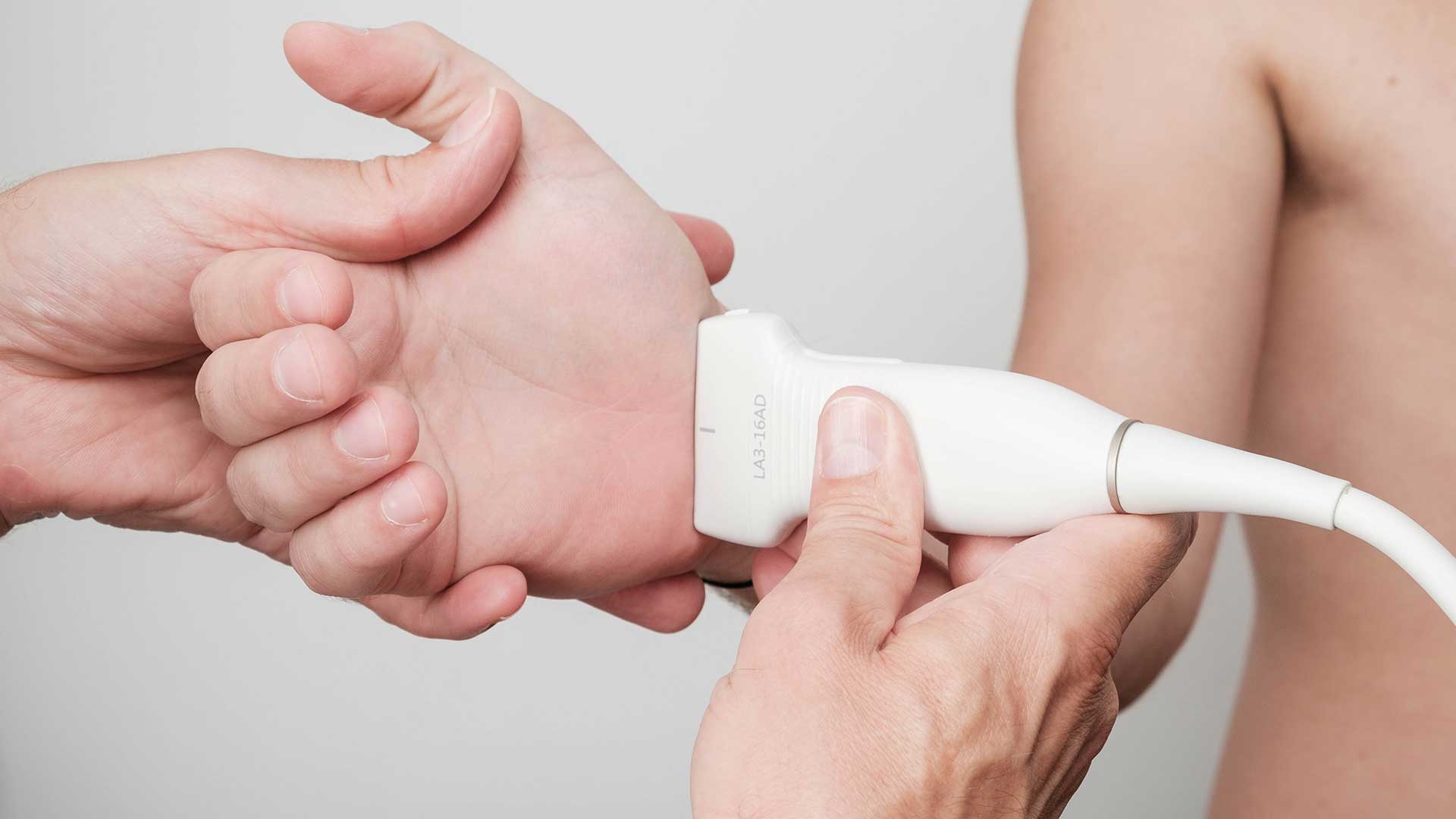
Ultrasound of peripheral nerve structures
NERVE COMPRESSION, NEUROPATHY, NERVE SHEATH TUMOUR
Diagnostic ultrasound of peripheral nerve structures is an integral part of soft tissue assessment, and with advances in ultrasound machines it is often the sole purpose of the examination.
Diagnostic ultrasound of the peripheral nerves and their sheaths makes it possible to assess superficial nerves, especially those running through superficial tunnels where these delicate structures can become compressed, which is why diagnostic ultrasound is a great help with compression neuropathies and with post-traumatic and postoperative conditions.
One of the most common compression neuropathies is carpal tunnel syndrome – it shows itself through tingling, weakness and pain in the hand and forearm, and through muscle wasting. Because it is quick and simple to use, diagnostic ultrasound is an effective and painless method for detecting neuromuscular disorders. Diagnostic ultrasound is increasingly used to identify problems before surgery, to establish the indication for surgery and for postoperative follow-up.
The method is painless, safe and repeatable, and it is also an excellent complement to electromyography with nerve conduction studies (EMNG). On the basis of clinical signs and the ultrasound image, some surgeons decide to operate, while for physiatrists and rheumatologists the ultrasound image allows better-targeted procedures, injections and other methods.


