
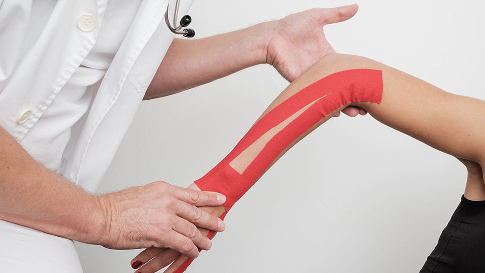
Elbow rehabilitation in sport
Elbow injuries make up a significant part of rehabilitation medicine, in children and adults alike. The anatomy here is distinctive: three bones articulate with one another, alongside vascular and nerve structures. Anatomically and functionally, the lower part of the humerus (the upper arm bone) belongs to the elbow. The distal humerus (the lower part of the upper arm bone) is exposed to fractures and dislocations; in particular, fractures or avulsions of the epicondyles, either medial or lateral, can occur. Fractures can be intra-articular or extra-articular. The second bone that makes up the elbow is the radius, which at its upper (proximal) end has a head in which rotation takes place. A fracture here can be intra-articular and multifragmentary, in which case rehabilitation is very complex and lengthy.
The third bone that makes up the elbow is the ulna. Fractures can affect its larger process, which we call the olecranon, and its smaller one, which we call the coronoid process. Such fractures prevent movement at the elbow, above all extension, and often also require surgical treatment. Elbow dislocations are particularly common in childhood. A Monteggia fracture is a fracture of the ulna combined with dislocation of the head of the radius.
Fractures of the shaft (diaphysis) of the forearm bones – one or both – occur in a fall on an outstretched hand or from a direct blow to the shaft of the bone, and they are the most common forearm fractures. Osteochondritis dissecans of the elbow presents with pain, reduced movement and swelling. Sports injuries are a common cause, and it is diagnosed through a medical examination that raises the suspicion and considers the differential diagnosis – a clinical examination and an X-ray, which we supplement with MRI. Soft-tissue injuries can affect the elbow area and the functionally linked areas of the upper arm, forearm and hand. Biceps tendon trauma occurs after a forceful impact, but also as a microtraumatic lesion caused by repetitive movements. The doctor's clinical examination, diagnostic ultrasound and magnetic resonance imaging are particularly important. The rehabilitation process will be adapted to each individual's needs. Injuries to the triceps muscle and tendon in the shoulder and elbow area may be combined with changes at the entheses and the olecranon.
The extensors and flexors of the forearm and hand attach to the elbow, that is, to the epicondyles: the common extensor origin is on the lateral epicondyle, while the common flexor origin is on the medial epicondyle. The elbow is also surrounded by ligaments – the medial and lateral collateral ligaments – while the head of the radius is encircled by the annular ligament, which is important for stability and for rotation of the forearm and hand at the elbow. Elbow tumours and infections will be covered in separate sections. Inflammatory joint diseases (arthritis, enthesitis) or degenerative changes of the elbow joint in osteoarthritis, secondary arthritis and other types of arthropathy can, apart from trauma, be part of metabolic conditions or the result of infection.
In sport, we pay particular attention to nerve entrapment syndromes at sites where nerve and vascular structures come into close contact with other structures, or lie superficially and are exposed to direct trauma. In the ulnar groove of the humerus, the ulnar nerve is exposed to injury, with symptoms that also appear distal to the site of injury, in the hand – in the 4th and 5th fingers. Apart from the carpal tunnel at the level of the wrist, the median nerve can be compressed in the upper forearm and elbow area. The radial nerve can also be injured anywhere along its course.
At our polyclinic, sports rehabilitation of the elbow is led by a physiatrist and a kinesiologist.
Appointments: 01 3737 333 · info@faktorzdravlje.com · Domobranska 25, Zagreb


