
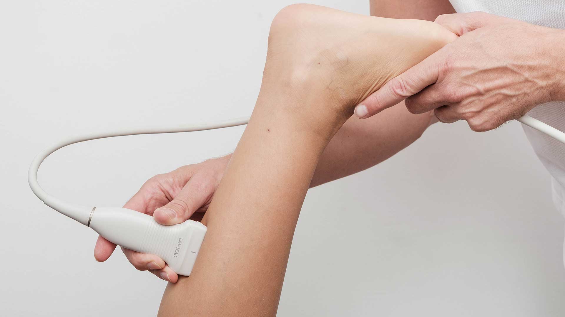
Muscle and tendon ultrasound
Diagnostic ultrasound is superior in assessing soft tissue damage, especially in superficial tissues. It correlates very well with magnetic resonance imaging, is cheaper and quicker, and can be repeated many times without risk to the patient. Ultrasound shows the thickness of a muscle and its atrophy, hypotrophy or hypertrophy. Traumatic injuries of muscles, tendons and ligaments are common in athletes, and timely diagnosis is very important to speed up healing. The same applies to contusions (bruises, small tears), ruptures (tears, loss of continuity), strains and sprains.
A haematoma (bruise) can appear as a small area of inhomogeneity, but a large cystic swelling that pushes other structures apart can also form. A seroma usually forms at the site of an older haematoma, but it can have other causes too. After a haematoma, heterotopic ossification can develop (e.g. myositis ossificans). Such changes involve deposits of mineralised tissue where there should be none. Muscle, tendon and ligament lesions (damage) usually end with a smaller or larger scar, which can limit function or cause pain. Other tests are a good complement to rule out other possible conditions.
Diagnostic ultrasound detects haematomas, fluid collections, effusions and cysts.
Diagnostic ultrasound can be used to monitor changes in lymph nodes, tissue infections and inflammation (abscesses, bursitis, arthritis, rheumatoid nodules, synovitis, tenosynovitis), erosions, osteophytes, cartilage changes in various diseases, superficial foreign bodies, lipomas, sebaceous cysts, nerve sheath tumours (in differential diagnosis), vascular anomalies, ganglion cysts, atypical superficial bone masses, exostoses and more.


